
|
|
|
Call our offices at: (310) 274-3481 (800) 964-0404
Beverly Hills
9301 Wilshire Boulevard
Suite 406A
Beverly Hills, California 90210

|
|
 |
|
|
INSURANCE & WORKERS' COMPENSATION
ACCEPTED!
We accept most types of
insurance
providers and specialize in the treatment of
workers' compensation injuries to the hand and upper extremity. |
|
|
|
Advanced Interventional Pain Management
Radiofrequency Neurotomy
Discography & Myelography
Discectomy and Microdiscectomy
Intradiscal Electrothermal Therapy (IDET)
Intrathecal Pump Implant
Spinal Cord Stimulation
Vertebroplasty
Our vertebral column, or spinal column, is made up of 33 individual bones that function to protect the spinal cord, nerve roots and internal organ; act as a base for attachment for ligaments, tendons and muscles; act as structural support for our head and body; and allows for flexibility and mobility. These individual bones, known as vertebrae, are composed of different structures including intervertebral discs and facet joints that contribute to the overall function of the spinal column. When a disc is ruptured or herniated, a facet joint is damaged, a vertebrae is fractured, or nerves are injured; advanced, minimally invasive procedures are required to repair, fix and alleviate pain emanating from these problems. For patients suffering from any of these injuries or conditions, the physicians at Avosant Surgical Associates will decide the proper treatment plan to ensure correction and relief, recommending surgical procedures only when necessary. Listed below are common minimally invasive procedures performed to correct and alleviate pain emanating from different injuries or damage to the spinal column.
Radiofrequency Neurotomy
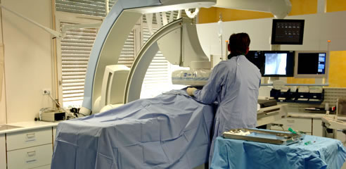
Radiofrequency neurotomy is a minimally invasive procedure used to relieve pain in the neck and back in the lumbar and cervical spine due to damaged facet joints by using a radiofrequency electrode to disrupt the medial branch nerves that send pain messages to the brain. A local anesthetic is used to numb the tissue all the way down to the spinal column. An x-ray devise known as a fluoroscope, is used to guide a needle like tube called a cannula to the irritated nerves. A radiofrequency electrode is inserted into the cannula and tests the position by sending a weak electric jolt. The electrode is then used to heat and cauterize the nerve to disrupt the pain signals to the brain, multiple nerves can be cauterized at one time if needed. The pain may increase for the first week after receiving a radiofrequency neurotomy, however, full relief will be felt within one month.
Discography & Myelography
Discography is a procedure used to diagnose and determine the cause of back pain and if it is caused by one or more discs. Discography pressurizes the discs with injection of a sterile fluid into the center of one or more discs. The patient is given IV medication to relax, however, they must be awake to communicate with the doctor about the pain felt during the procedure. A local anesthetic is used to numb the skin all the way down to the spine. An x-ray devise known as a fluoroscope, is used to guide the needle to the edge of the disc/s, a smaller needle is then guided through the other and is inserted into the middle of the disc. The disc is then pressurized with injection of a contrast die, this can be done to more than one disc at a time. The patient will then feel pressure or pain which they will describe to the doctor. An image is taken of the disc/s with the fluoroscope. If is the same pain the patient feels normally, this may indicate a diseased disc and may need to undergo further testing such as a CT scan. After the procedure the patient may feel soreness and discomfort. It is advised for patient to take an anti-inflammatory medication such as IB profen and ice the area until the soreness goes away.
Myelography is a diagnostic procedure used to identify spine, spinal cord, and nerve root problems. Myelography uses an x-ray sensitive contrast die that is injected into the spinal canal in order to provide clearer x-ray or CT scan images to make identification of pinched or damaged nerves easier. Myelography is an outpatient procedure. A local anesthetic is used to numb the skin and tissue of the lower back down to the spinal column. A needle is guided into the spinal column and through the dura, the special x-ray sensitive contrast die is then injected and mixes with the spinal fluid allowing it to flow through the spinal canal. The spine is then studied with an x-ray or CT scan providing clear images for identification and diagnosis. The contrast die can either be left in the spinal column and will be absorbed by the body, or it can be withdrawn by the doctor.
Discectomy and Microdiscectomy
A discectomy and microdiscectomy procedure is used to alleviate pain caused by herniated discs.
A microdiscectomy is a minimally invasive procedure that uses an endoscope or surgical microscope through a very small incision to see and remove portions of herniated discs that protrude out of the disc space putting pressure on the spinal cord and/or nerves. The patient is put under general anesthesia. The surgeon uses an x-ray devise called a fluoroscope to confirm the position of the disc. A small incision is made in the back near the affected discs and microsurgical instruments are inserted. A hole is made in the vertebrae, or small piece of bone may be removed, in order to properly view and reach the spinal canal and nerve roots. The surgeon uses the endoscope to view and inspect the herniated disc and is able to remove the degenerate or herniated portion of the disc nucleus to relieve the pressure on the nerve roots. A laser and radiofrequency probe is used to treat the disc wall and prevent further leakage. The patient may need 1 day of bed rest and can return to normal activities in 1-6 weeks.
A discectomy is a surgical procedure that removes some of the nucleus of an intervertebral disc which allows the disc to reabsorb the herniation. The exact technique varies and is dependent on the location of the herniated disc. Generally, it is a minimally invasive procedure and is done under local anesthetic. This procedure uses a small needle, called a cannula, and a probe devise. The cannula is inserted into the herniated disc and is guided by an x-ray devise called a fluoroscope. The probe is then inserted through the cannula where it is able to remove a small portion of the disc nucleus allowing for reabsorption of the herniation. After the procedure, the patient may need a day of best rest and possibly physical therapy and may return to normal activity within 1-6 weeks.
Intradiscal Electrothermal Therapy (IDET)
Intradiscal electrothermal therapy is a minimally invasive surgical procedure that is used to alleviate low back pain cause by diseased discs or small disc herniations. The procedure is done under local anesthesia and a mild sedative is used to relax and calm the patient. An x-ray devise known as a fluoroscope, is used to guide a hollow needle into the disc. A electrothermal catheter or heating wire is inserted through the hollow needle and maneuvered to find the affected area. The wire is then slowly heated to approximately 195 degrees Fahrenheit, heating the disc wall. This heat shrinks and repairs any tears in the disc wall, small nerve endings may also be cauterized in order to make them less sensitive. The patient may feel pain, however this indicates that the heat is being applied to the appropriate area. The instruments are removed and the patient is allowed to go home the same day.
Intrathecal Pump Implant
An intrathecal pump implant is used to relieve chronic pain in patients in which conservative procedures have failed and surgery is unlikely to help. The intrathecal pump dispenses small amounts of medication in the intrathecal space, the space around the spinal cord, to prevent pain signals from going to the brain. For the first surgery, the trial procedure, local anesthetic is administered and a catheter is inserted into the intrathecal space that is connected to a temporary pump to see if the pump successfully relieves the pain. If the trial is successful, a second surgery is required to permanently implant the pump. The patient is put under general anesthesia. The temporary catheter is removed and a permanent catheter is implanted, and the pump is implanted under the skin, generally in the abdomen. The pump battery lasts between 3-5 years and a new pump will need to be implanted. The medication is administered and controlled using an external program. Regular doctor visits are required in order to refill the pump medication. After surgery, the patient may feel mild discomfort and swelling at the surgical site. Over time, the pump may move and may require additional surgery to reposition or replace the pump.
Spinal Cord Stimulation
 Spinal cord stimulation is a procedure that uses electrical impulse to relieve chronic pain in the back, arms, and legs caused by neuropathic pain by preventing pain signals from reaching the brain. This procedure is performed on patients where conservative treatments have failed. Spinal cord stimulation requires a trial period to see if the procedure works. For the first surgery, a local anesthesia is used to numb the incision site. Through an epidural needle, one or more insulated wire leads are guide into the epidural space, the area around the spinal cord. Electrodes at the end of the leads produce electrical pulses that block the pain. For the next week, the patient will give thorough feedback to the doctor to determine if spinal cord stimulation works. If the patient and doctor determine that spinal cord stimulation provided significant relief, a permanent implant will be administered. For this surgery, the patient may be sedated or put under general anesthesia. The insulated wire leads are inserted into the predetermined locations in the epidural space. A small incision is made and the pulse generator battery is implanted under the skin, generally in the buttocks or abdomen, and is programmed with an external control unit that the patient can use to turn the system on or off, switch programs, and adjust the stimulator power levels. After surgery, the patient may experience mild discomfort or swelling at the incision site.
Vertebroplasty
A vertebroplasty is a minimally invasive procedure used to repair spinal fractures caused by osteoporosis and cancer by stabilizing the spine to provide rapid pain relief and to prevent further weakening of the vertebral body. During a vertebroplasty, the area on the back is numbed with local anesthetic and a tube is inserted through a small incision and is guided into place with an x-ray devise known as a fluoroscope. One or more needles are inserted through the tube and into the collapsed vertebra to collect bone tissue for study. The weakened area is then filled with bone cement creating an internal cast to stabilize the vertebra body. The instruments are removed and the patient is allowed to go home the same day.
Top of the Page |
|